- Shop
- Learn
-
-
- Buy The Sunflower Now
Feel the difference
Get Started with JustLight
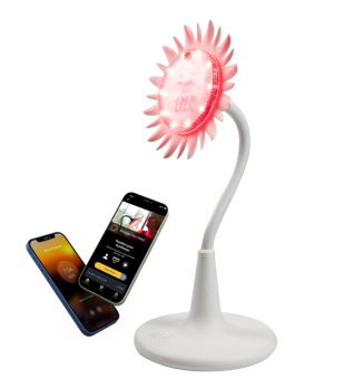
Sunflower is the most advanced light therapy available today.

-
- Collaborate
- Blog
- Quiz
- My Account
Free Shipping in the 48 States!
The science of photobiomodulation (PBM), also known as low-level laser therapy (LLLT), continues to shed light on various biological effects,...
The science of photobiomodulation (PBM), also known as low-level laser therapy (LLLT), continues to shed light on various biological effects, including cellular responses and dose-response relationships. This fascinating field has advanced through research exploring the biphasic dose-response curve, where both low and high doses of light can produce suboptimal effects, while optimal doses yield maximum biological responses. PBM, using specific light wavelengths, is gaining traction for its diverse applications, such as reducing inflammation, promoting wound healing, and even supporting cognitive health.
At the core of PBM research is the biphasic dose-response curve, which describes a hormetic effect—the idea that small doses of light can stimulate a beneficial response, while larger doses can lead to inhibitory effects or adverse reactions. The dose-response curve helps clarify how different dose levels interact with biological systems to either promote or inhibit specific functions. This principle is critical in PBM therapy because it determines the effective doses needed to stimulate cellular repair while avoiding overexposure that may hinder healing.
The biphasic dose-response curve is often illustrated as a U-shaped or bell-shaped curve, representing how both insufficient and excessive doses result in reduced effectiveness, with a middle optimal dose providing the most positive outcomes. This model has been observed across multiple biological functions, including collagen production, cell proliferation, and tissue repair in response to PBM treatment. Various mathematical models, such as the Brain-Cousens model, have been developed to predict the effects of PBM under different dose conditions, with the goal of optimizing treatment parameters.
PBM exerts its therapeutic effects at the cellular level by influencing mitochondrial function and increasing the production of adenosine triphosphate (ATP), the energy currency of the cell. The hormetic effects observed in Photobiomodulation therapy mean that low levels of light can promote cell health and cellular regeneration, while higher doses might lead to inhibitory effects such as oxidative stress and impaired cellular repair.
Low-level laser irradiation has been shown to stimulate endothelial cells and other cellular structures, increasing blood circulation and facilitating wound healing. Studies have observed these biphasic responses in various cell types, including MCF-7 cells (a breast cancer cell line), which have demonstrated enhanced repair at moderate doses of light but adverse reactions at high levels. The Brain-Cousens and Cedergreen equations help researchers model and understand these biphasic effects across different biological systems.
A growing body of evidence highlights the potential of PBM in treating chronic inflammation and neurodegenerative diseases. By targeting the mitochondria, PBM reduces inflammation at the cellular level while supporting neurogenesis and tissue repair in damaged regions. Experimental datasets and hormetic dose-response curves have demonstrated PBM’s ability to modulate both inflammatory markers and cognitive performance.
In particular, PBM has been shown to improve outcomes in individuals with traumatic brain injuries (TBI) and neurodegenerative disorders such as Alzheimer’s and Parkinson’s disease. The therapeutic wavelengths used in these treatments often range from 600 to 900 nm, penetrating deep into tissues and promoting cellular regeneration. Recent studies have also explored PBM’s potential to alleviate symptoms associated with psychiatric disorders, such as depressive disorder and anxiety, suggesting that the therapy may have significant mental health benefits as well.
A key challenge in advancing PBM is identifying the correct dose for each condition. Studies have consistently demonstrated biphasic effects, meaning that finding the right balance of light exposure is crucial to achieving therapeutic outcomes. Clinical studies have reported that moderate doses of near-infrared light and red light offer the most beneficial effects, with excessive doses leading to diminished outcomes or even negative impacts.
For instance, research by Yang et al. and Salehpour et al. has explored PBM’s role in reducing brain swelling and supporting recovery in cases of closed-head TBI. These studies have highlighted the importance of transcranial Photobiomodulation (tPBM) in mitigating axonal injury and promoting cognitive recovery. Photomed Laser Surg publications have also discussed the importance of accurate dosing in order to achieve optimal therapeutic benefits without the risk of overexposure.
While PBM is gaining recognition as an effective treatment for various conditions, from chronic skin issues to neurodegenerative diseases, more research is needed to refine the understanding of hormetic dose responses. Future studies will likely focus on further exploring the biphasic dose-response curve, optimizing treatment doses, and assessing PBM’s applications in a broader range of neurological conditions. Additionally, as more light therapy devices are developed, understanding the power output, wavelengths of light, and optimal treatment protocols will be critical to delivering the best results.
JustLight’s Sunflower, an advanced PBM device, stands out for its ability to deliver targeted therapy at various light wavelengths, ensuring that users receive the appropriate dose for their condition. This makes it a versatile and valuable tool in a wide array of light therapy treatments. As PBM technology advances, innovations like the Sunflower will continue to play a pivotal role in revolutionizing healthcare.
The benefits of red light therapy💡
Have questions about Sunflower? Send us a DM! 📩
#redlighttherapy #redlighttherapybenefits

Here’s your sign to give Sunflower a try🌻 so many benefits in just 1 device 👏
Use the code LIGHT99 for $99 off your first order 🤩
.
.
Have questions? Send us a DM or click the link in our bio to visit our website!

Take advantage of nature’s healing power..LIGHT! 💡
Most of us don’t have access to the sun on a daily basis depending on our jobs, location, and lifestyle. Sunflower by JustLight provides an easy user-experience to incorporate light into your routine regardless of where you are🌻
Have questions? Send us a DM or click the link in our bio 📩

Unbox Sunflower with us!🌻
1. Download our app (Sunflower by JustLight)
2. Remove eyewear from plastic bag
3. Remove power cord
4. Plug into wall and device to turn on the device
5. Select the routine of your choice (11 different options!)
Have questions? Send us a DM or use the link in our bio to send us an email 📩

This before & after transformation makes us grateful for Smart Light Therapy! The only technology that can accurately dose the correct amount of light to reach deep layers of your skin.
Sick of your wellness routine not providing the results you’re looking for? Switch to Sunflower by JustLight🌻
Click the link in our bio to learn more or use code LIGHT30 for 30% off your order 👏

Thank you to everyone who made it to our first wellness event! We’re excited to announce our product, Sunflower, will be part of yoga/meditation classes located at the Canvas 3.0 gallery in the Oculus (NYC) starting this month. Stay tuned for more updates 🙌 We can’t wait for everyone to be able to experience this amazing class!
Classes by @a_cuffs 🧘♀️
Studio space by @blakelee_mover ✨
Wellness event @hellojustlight x @togeth3r 🍃

Symptoms weighing you down? It’s time to find relief 😌
Sunflower can improve:
-Energy
-Mood
-Pain
-Hair growth
-Sleep
-Skin
-Blood circulation
-Cognitive function
& much more 👏
Click the link in our bio to learn more about Sunflower by JustLight or send us a DM 📩

Pilates 🤝 JustLight
How does our product benefit your workouts? 🤔
Red and near-infrared light heal damaged muscle tissues and increase blood flow to your muscles. Our muscle routine is great to warm up muscles before an intense workout or to recover post-workout 💪
Have questions? Send us a DM! 📩
Have questions? Send us a DM!

Calling all LA wellness lovers! JustLight will be available at @bspokela for the rest of this month located at 439 N Fairfax Ave 🌻
Stop by and experience the healing effects of Sunflower by JustLight💡
